The cleft palate, when isolated, is not visible from the outside. This is why sometimes the diagnosis may not be done immediately at birth. It is due to a defect of fusion between the two palatal blades towards the 40th day of the embryonic development. The cleft can affect the whole palate, it is then a velo-palatal cleft, or only the most posterior part of the palate (photo 1), it is then a velar cleft.
The cleft palate can let food pass from the mouth to the nose. Breastfeeding is difficult, usually inefficient because the child sucks air through the nose. It is advisable to use a suitable feeding bottle (see Treatment The palate). There are particular forms of cleft palate, round in front (photo 2), they evoke a Pierre Robin syndrome (see Treatment Pierre Robin Syndrome).
Sometimes the velar cleft is incomplete, there can still be some mucosa in the middle, but there is no muscle, it is a cleft under the veil’s mucosa (photo 3). Sometimes in cleft palates, the Eustachian tube does not work properly, this is responsible for serous otitis. The installation of tympanostomy tubes is then necessary (see The ear).
After the surgery, carried out between 4 and 6 months old, a surveillance of the speech and the growth of the maxilla is essential. An early orthophonic assessment and regular follow-ups are performed, if necessary speech therapy is recommended (see Speech). Maxillary growth is controlled, early orthodontic treatment can be organised to stimulate growth (see Teeth). If the speech is incorrect between 4 and 6 years, despite good speech therapy, a second surgery is proposed, it is a pharyngoplasty. It helps correct air leaks through the nose during speech.
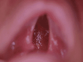
Cleft palate bike isolated
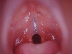
Cleft mucosa of the veil
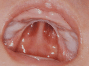
Cleft palate in Roman arch (robin stone syndrome)